A stubborn ear infection is solved by a chain of tests, not a bottle of drops, because each test answers a question the others can’t. Cytology, the quick swab-and-slide exam, identifies whether yeast, bacteria, or mites are living in the canal. Culture and sensitivity testing names the exact organism and the drug that will beat it when the bacteria turn out to be resistant. And biopsy answers the question when it’s not an organism at fault, like growths, glandular diseases, or cysts. Ears treated without those answers tend to calm down just long enough to relapse, often harder to clear each time.
Cupertino Animal Hospital has built a dedicated Pet Ear Care Center around exactly these cases, offering advanced ear care for dogs and cats where the workup matches the problem instead of restarting the same treatment. Cytology and culture tell us what is growing, our 4K video otoscope shows the deep canal and eardrum in remarkable detail while we collect samples and biopsies precisely, and 3D cone beam CT reveals the middle and inner ear that no scope can reach. If your pet’s scratching and head shaking keep coming back, book an exam and we will run down the answer step by step.
Reading the Ear Instead of Guessing at It
- Ear cytology identifies which organism is actually in the canal, because yeast, bacterial, and mite infections look nearly identical from the outside yet each needs a different medication.
- Rod-shaped bacteria on the slide, especially Pseudomonas, change the whole plan: they signal a resistant infection that calls for culture and sensitivity testing before the next prescription.
- Not every chronic ear is an infection at all; polyps, masses, and middle-ear disease mimic one, and biopsy and advanced imaging are how they get found.
- A recheck cytology confirms the organisms are truly gone before medication stops, which is one of the best ways to keep an ear problem from turning chronic.
What Exactly Is Ear Cytology?
When your vet swabs the ear and slips away to the microscope, that’s ear cytology in action: the debris goes onto a slide with a stain that makes yeast, bacteria, mites, and cells stand out clearly under magnification. The whole thing is fast and gentle, and most pets barely register that it happened.
The ear canal makes cytology especially valuable because of its shape. The canine and feline canal takes an L-shaped bend before reaching the eardrum, trapping warmth and moisture in a chamber air struggles to reach, which is why otitis externa, inflammation of the outer canal, is one of the most common problems in veterinary medicine. Whenever the canal has inflammation, the organisms that normally live there in limited numbers have a chance to multiply and cause a secondary infection.
Because this is one of our in-house diagnostics, the slide gets read right here instead of being shipped to an outside lab, so we can talk through what we find while your pet is still in the room, with the plan built around real information rather than a hunch.
What Does the Vet Actually See on the Slide?
Under the microscope, a stained ear sample tells a detailed story. We identify and count what’s there, because both the organism and its numbers decide the treatment: a few yeast per field can be normal, while sheets of them confirm an overgrowth that needs therapy.
| What shows up on the slide | What it usually is | What it means for treatment |
| Budding, peanut-shaped yeast | Malassezia, a normal resident grown out of control | Antifungal therapy plus managing the trigger that let it overgrow |
| Round bacteria (cocci) | Most often Staphylococcus, sometimes Streptococcus | Usually responds to standard topical antibiotics |
| Rod-shaped bacteria | Often Pseudomonas or similar gram-negative bacteria | A resistance red flag; culture before choosing the next antibiotic |
| Mites or mite eggs | Otodectes, the common ear mite | A parasite treatment; antibiotics and antifungals do nothing |
| Sheets of white blood cells | Active, deeper inflammation | Points toward middle-ear disease or an immune-driven process |
A few of these findings deserve a closer look, because they change the plan the moment they appear.
Malassezia yeast live in small numbers in most healthy ears. Trouble starts when allergy, moisture, or a waxy canal lets them multiply, and the slide shows the difference between a resident population and a full overgrowth. That’s also why yeast infections relapse when only the yeast is treated: the underlying trigger reopens the door every time.
Cocci are the everyday bacterial finding, and most respond well to the usual topical medications, which is exactly why confirming them matters. An ear with simple cocci doesn’t need the big-gun antibiotics, and saving those drugs protects their power for the ears that truly need them.
Rods are the finding that stops us in our tracks. Pseudomonas in particular is the most feared organism in chronic ear disease: it thrives in damaged, moist canals, builds a slimy biofilm that shields it from medication, ulcerates the canal until it’s genuinely painful, and shrugs off many common antibiotics. Rods on the slide, especially alongside white blood cells, usually mean a deeper infection, possibly a ruptured eardrum, and always a culture before the next prescription.
Reading those patterns well takes real practice, and our veterinary team can tell the difference between a simple yeast flare and the rods that call for a much closer look.
When Is a Culture Needed Instead of Just Cytology?
Cytology tells us what kind of organism is present; it can’t tell us which drug will beat it. Culture and sensitivity testing answers that second question by growing the organism in a lab and testing it against a panel of antibiotics, so the next prescription is chosen by evidence rather than by another educated guess.
We reach for a culture in a handful of situations:
- Rods on the slide
- An ear that has failed a reasonable course of treatment
- A relapse soon after finishing medication
- Infection near a ruptured eardrum, where the wrong drug can damage hearing and balance
The sensitivity report matters most with Pseudomonas, where resistance to multiple drug families is common and the difference between the right and wrong choice is the difference between resolution and another six months of misery. Interpreting that report correctly is part of the advanced ear-infection workups our Pet Ear Care Center runs every week.
What If the Problem Isn’t an Infection at All? When Biopsy Comes In
Some “ear infections” that never clear turn out not to be infections. A growth in the canal traps wax and moisture, infection layers on top, and every treatment round clears the surface while the real problem sits underneath. Biopsy, taking a small piece of tissue for a pathologist, is how those cases finally get named.
The list of impostors is longer than most families expect, and these are just a few of the options:
- Ear polyps are benign growths, especially in cats, that arise from the middle ear or throat and block the canal from the inside.
- Ceruminous gland tumors, growths of the wax-producing glands, range from benign to malignant, and only histopathology tells them apart.
- Glandular hyperplasia thickens and narrows the canal until debris can’t escape.
- Cholesteatoma, a destructive keratin-filled cyst in the middle ear, quietly erodes surrounding bone while masquerading as one more stubborn infection.
Using video otoscopy with our advanced 4K endoscope, we can see these growths clearly, biopsy them precisely, and often remove polyps and foreign material like foxtails in the same procedure. The biopsy result then drives everything that follows: a benign polyp is removed and done, while a malignant tumor gets imaging to map its extent before surgery is planned.
How Do a 4K Endoscope and 3D Cone Beam CT Change the Diagnosis?
Endoscopy and CT let us see what a handheld otoscope never will. A standard ear exam views the first stretch of the canal through a cone of light. Our 4K video otoscope travels the full canal in high resolution with 100x better clarity than traditional equipment, showing the eardrum, subtle masses, ulceration, and retained debris on a screen we can walk you through in the exam room. Deep cleaning, flushing, and sample collection all happen under that same magnified view, so nothing is done blind.
Why Does Seeing the Eardrum Matter?
The eardrum is the fork in the road, and seeing it clearly changes treatment on the spot. An intact eardrum means the full range of topical medications is safe. A ruptured one rules several of them out and tells us the middle ear is likely involved. And myringitis, inflammation of the eardrum itself, is nearly impossible to identify without magnified visualization.
What’s Beyond the Eardrum?
Beyond the eardrum sits territory no scope can reach, and that’s where 3D cone beam CT takes over. It images the middle ear and the bulla, the bony chamber behind the eardrum, revealing the fluid and thickening of middle-ear infection, the bone changes of cholesteatoma, Eustachian tube disorders, and how far a tumor extends. That picture decides between continued medical treatment, a deep flush under anesthesia, or surgery, and it’s the reason complex ears referred to our Pet Ear Care Center leave with an actual diagnosis instead of a fifth bottle of drops.
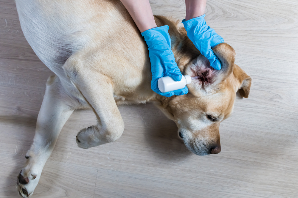
Frequently Asked Questions About Ear Cytology and Testing
Does the ear swab hurt my dog or cat?
For most pets, no. We use a soft cotton swab to collect a little debris from the canal, which takes only a few seconds and doesn’t require sedation. A pet whose ear is already very sore or badly inflamed may flinch, and in those cases we go slowly and, if needed, calm things with a little pain relief first. That quick swab spares your pet the far greater discomfort of a mistreated, lingering infection.
Can I treat my pet’s ear at home with leftover medication?
It’s tempting, but it often backfires. Leftover drops were matched to a past infection, and this one may be caused by an entirely different organism, so the old medication can do nothing while the real problem worsens. Certain ear medications are also unsafe if the eardrum has ruptured, something you can’t check at home. A fresh swab and slide tell us what’s in there now, so hold off and let us take a quick look first.
My pet has been treated for the same ear infection four times. What now?
An ear that relapses that often needs a different kind of workup, not a fifth round of the same drops. That usually means cytology plus a culture to check for resistant bacteria, a 4K otoscopic exam to inspect the deep canal and eardrum for growths or damage, and sometimes cone beam CT to look behind the eardrum for middle-ear disease. Somewhere in that sequence, the reason the infections keep returning almost always shows itself.
Healthy Ears Start With a Look Inside
Ear cytology isn’t an extra step tacked onto the visit. It’s the difference between guessing and knowing, and it’s the first link in a diagnostic chain, from slide to culture to biopsy to imaging, that can solve even the ears that have failed treatment for years. If your pet is scratching, shaking, or holding an ear low, schedule a visit with the ear experts at Cupertino Animal Hospital, and we’ll find a real answer.